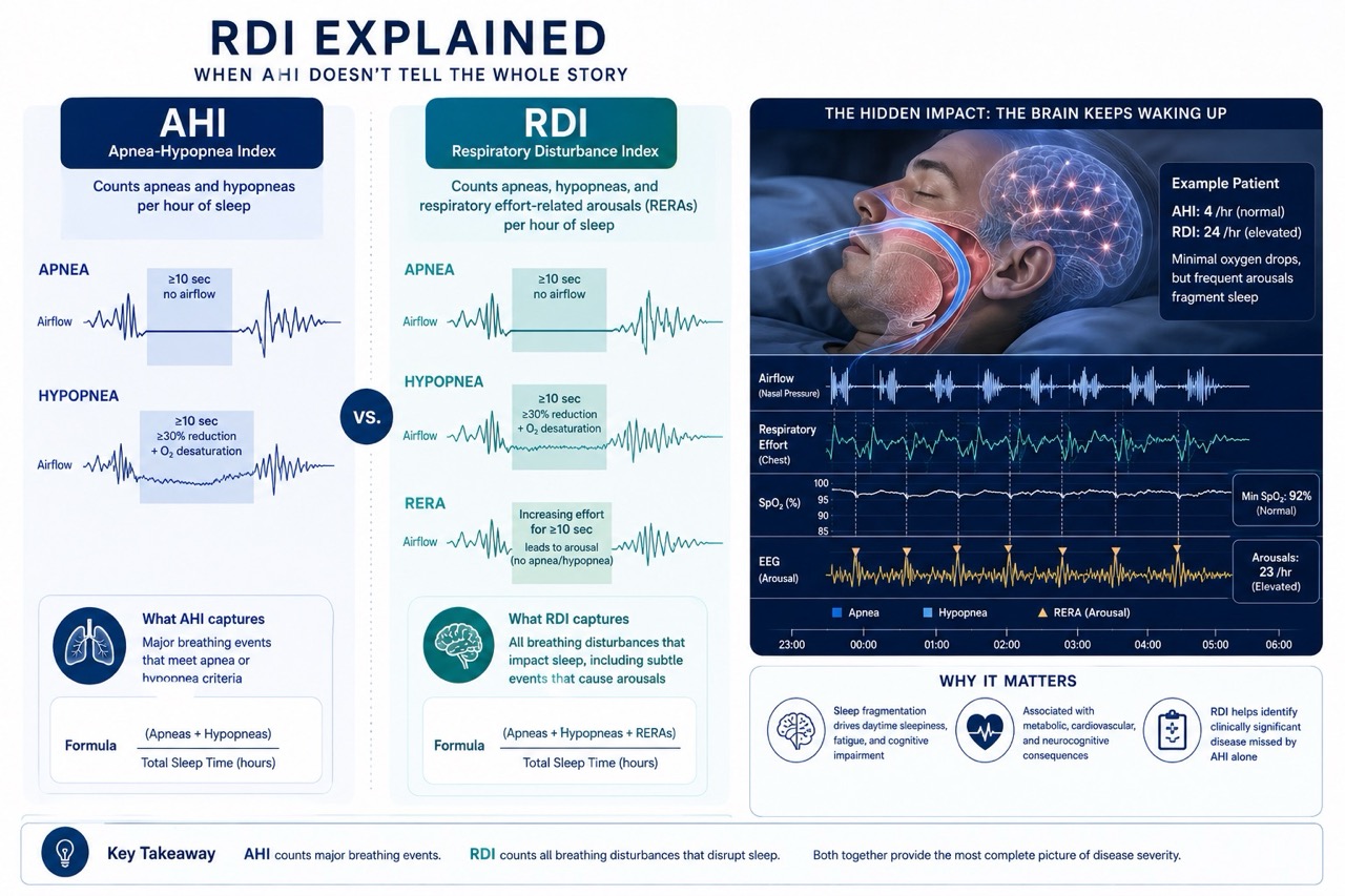
Understanding respiratory disturbance index (RDI): when AHI doesn’t tell the whole story
Most patients undergoing a sleep study become familiar with one number above all others: the Apnea-Hypopnea Index (AHI).
AHI serves as the foundation of sleep apnea diagnosis and treatment decisions throughout much of the world. However, many sleep reports contain another metric that often receives far less attention: the Respiratory Disturbance Index (RDI).
In some cases, patients may have a normal or only mildly elevated AHI but a substantially higher RDI. These individuals often experience significant symptoms despite appearing to have relatively mild sleep-disordered breathing according to traditional AHI criteria.
Understanding RDI helps explain why some patients remain exhausted, sleepy, or cognitively impaired even when their AHI appears reassuring.
What is RDI?
The Respiratory Disturbance Index measures the average number of respiratory disturbances occurring per hour of sleep.
Unlike AHI, which includes only apneas and hypopneas, RDI also incorporates respiratory effort-related arousals (RERAs).
The formula is:
RDI = (Apneas + Hypopneas + RERAs) ÷ Total Sleep Hours
Because it captures additional respiratory events, RDI is always equal to or higher than AHI.
Understanding the components
Apneas
Complete or near-complete cessation of airflow lasting at least 10 seconds.
Hypopneas
Partial reductions in airflow associated with oxygen desaturation or arousal.
RERAs
Respiratory effort-related arousals that do not meet criteria for apnea or hypopnea but still disrupt sleep.
These events are often the reason RDI exceeds AHI.
What is a RERA?
A respiratory effort-related arousal is a subtle breathing disturbance characterized by:
- Progressive airflow limitation
- Increasing respiratory effort
- Brief cortical arousal from sleep
without meeting formal scoring criteria for apnea or hypopnea.
In other words, breathing becomes difficult enough to wake the brain, but not severe enough to qualify as a conventional respiratory event.
The patient may never remember waking.
Nevertheless, hundreds of these disruptions can fragment sleep architecture.
Why RDI can be significantly higher than AHI
Consider two patients.
Patient A
AHI = 5
RDI = 6
Minimal difference.
Patient B
AHI = 5
RDI = 22
Numerous respiratory effort-related arousals occur throughout the night.
Traditional AHI interpretation might suggest mild disease.
RDI suggests substantial sleep disruption.
These patients often report severe daytime symptoms despite relatively low AHI values.
Why sleep fragmentation matters
Sleep is not restorative simply because a person remains unconscious.
Normal sleep requires:
- Stable sleep architecture
- Continuous sleep stages
- Minimal arousal activity
Repeated respiratory arousals interrupt this process.
Consequences include:
- Excessive daytime sleepiness
- Fatigue
- Poor concentration
- Memory impairment
- Mood disturbances
- Reduced quality of life
In many patients, sleep fragmentation may contribute more to symptoms than oxygen desaturation.
RDI and upper airway resistance syndrome (UARS)
One of the most important applications of RDI involves Upper Airway Resistance Syndrome (UARS).
Patients with UARS often demonstrate:
- Normal or near-normal AHI
- Minimal oxygen desaturation
- Frequent respiratory effort-related arousals
- Significant daytime symptoms
Without RDI assessment, these patients may be incorrectly told their sleep study is normal.
RDI provides a more complete picture of respiratory-related sleep disruption.
Why AHI can underestimate disease burden
AHI focuses primarily on:
- Airflow reduction
- Oxygen desaturation
RDI expands assessment by considering:
- Increased respiratory effort
- Sleep fragmentation
- Subclinical airway obstruction
As a result, RDI often identifies clinically important disease that may not be reflected by AHI alone.
This is particularly relevant in:
- Younger patients
- Women
- Lean individuals
- UARS patients
- Patients with persistent fatigue despite low AHI
How RDI is measured
Accurate RDI measurement generally requires polysomnography.
Identification of RERAs often depends upon:
- Nasal pressure transducers
- Respiratory effort monitoring
- EEG arousal detection
Many home sleep apnea tests cannot reliably detect RERAs.
Consequently, home testing may underestimate sleep disruption in certain patients.
RDI versus AHI
| Metric | AHI | RDI |
|---|---|---|
| Apneas included | Yes | Yes |
| Hypopneas included | Yes | Yes |
| RERAs included | No | Yes |
| Measures sleep fragmentation | Limited | Better |
| Captures subtle airway resistance | No | Yes |
| Useful in UARS | Limited | Excellent |
Clinical implications
When evaluating sleep study results, clinicians should consider both AHI and RDI.
A low AHI does not always indicate an absence of clinically meaningful disease.
Patients with elevated RDI may experience:
- Significant daytime impairment
- Reduced sleep quality
- Cardiovascular stress
- Persistent symptoms
despite apparently reassuring AHI values.
Understanding this distinction helps avoid underdiagnosis and undertreatment.
The future of respiratory event assessment
Modern sleep medicine increasingly recognizes that respiratory disease exists along a spectrum.
Traditional classifications based solely on AHI may fail to capture:
- Flow limitation
- Respiratory effort
- Sleep fragmentation
- Arousal burden
Metrics such as RDI provide a broader view of how breathing abnormalities affect sleep quality and overall health.
As the field moves toward precision medicine, comprehensive assessment will likely rely on multiple complementary metrics rather than a single number.
Key takeaways
- RDI includes apneas, hypopneas, and respiratory effort-related arousals.
- RDI is always equal to or higher than AHI.
- Elevated RDI may explain symptoms in patients with normal or mild AHI.
- RERAs can significantly fragment sleep despite causing little oxygen desaturation.
- RDI is particularly important in Upper Airway Resistance Syndrome.
- Evaluating both AHI and RDI provides a more complete understanding of sleep-disordered breathing.
References
- American Academy of Sleep Medicine (AASM) Manual for the Scoring of Sleep and Associated Events.
- Guilleminault C. Upper Airway Resistance Syndrome.
- Malhotra A, Ayappa I, Ayas N, et al. Metrics of Sleep Apnea Severity Beyond the Apnea-Hypopnea Index.
- Eckert DJ. Phenotypic Approaches to Obstructive Sleep Apnea.